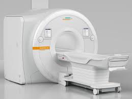
Magnetic resonance imaging, also known as MRI, is one of the most advanced imaging technologies and is widely used in medicine today. Since it was developed in the 1970s, MRI has altered diagnostic processes, especially in neurology, cardiology, and oncology. This article covers MRI’s underlying principles, its clinical applications, and its future prospects.
How MRI works
MRI uses the magnetic properties of atoms within our body to generate detailed images of internal structures. It is exceptionally good at picturing soft tissues. Most MRI machines rely on the behavior of hydrogen atoms, which are abundant in human tissue due to their presence in water and fat molecules.
The MRI machine creates a powerful magnetic field that temporarily aligns tiny particles in our body’s hydrogen atoms, which -as we mentioned earlier- are abundant because we’re made mostly of water. After that, a quick pulse of radio waves nudges these particles out of alignment. When they settle back, they give off energy, which the MRI machine picks up and uses to create detailed images of our body.
Two main types of signals, T1 and T2, play a key role in MRI imaging. T1 signals show how fast the particles realign with the magnetic field, while T2 signals reflect how quickly they lose synchronization. These slight differences help the MRI distinguish between various tissues, allowing for evident and accurate images.
Advantages and Limitations of MRI
One of the biggest advantages of MRI is its ability to generate highly detailed images without ionizing radiation. This makes it a safer option compared to CT scans and X-rays, especially for repeated use. MRI is efficient for imaging soft tissues, such as the brain, muscles, and internal organs, making it essential in many clinical settings.
However, MRI has some limitations. The machines are costly, both to purchase and maintain, making scans expensive for patients. MRI scans also take more time than other imaging methods, and patients must remain still for precise images, which can be exhausting for those with anxiety or claustrophobia. MRI is also unsuitable for patients with metal implants or pacemakers, as the magnetic field can interfere with these devices.
MRI in clinical practice
MRI plays a vital role in diagnosing and monitoring many health conditions. Here’s how it’s used across different fields of medicine:
Brain and Spine (Neurology): MRI is incredibly valuable for understanding what’s going on in the brain and spinal cord. Doctors rely on it to detect issues like brain tumours, strokes, and conditions. Because MRI can show detailed differences between types of brain tissue (like gray and white matter), it helps reveal even subtle abnormalities, making it a go-to tool for neurological problems.
Heart Health (Cardiology): For heart conditions, MRI helps doctors assess the heart’s structure and function. It can detect diseases in the heart muscle, check on heart valves, and see how well blood flows through the arteries. Cardiac MRI is especially useful for people who need detailed heart images but need to avoid radiation-based scans.
Cancer Detection (Oncology): MRI is often used to detect cancer early and begin treatment accordingly. It is particularly effective for examining soft tissue cancers like those in the breast, prostate, and liver. MRI can show the difference between cancerous and healthy tissue, helping doctors pinpoint where a tumor is and plan the best treatment path.
Muscles and Joints (Musculoskeletal System): MRI is crucial for injuries or disorders affecting muscles, ligaments, or joints. It is frequently used in sports medicine to diagnose torn ligaments, muscle injuries, and joint problems, providing detailed images that are crucial for proper treatment.
Abdomen and Organs: MRI can reveal problems with internal organs like the liver, pancreas, and kidneys, such as liver disease, tumors, or inflammation. With the help of special contrast agents, MRI can show detailed views of the abdominal organs, making it easier for doctors to monitor or diagnose conditions in these areas.
Technological Advances in MRI
Over the past few years, MRI technology has advanced significantly, making it faster and more precise:
High-Field Strength MRI: New MRI machines with field strengths of 3 Tesla and higher generate images faster and more detailedly. These high-field machines are particularly beneficial for studying complex structures such as brain tissue and small lesions, which were difficult to detect with lower field strengths.
Functional MRI (fMRI): fMRI is a specialized type of MRI that measures brain activity by detecting changes in blood flow. It has been a “quantum leap” in neuroscience, allowing researchers and clinicians to observe brain function in real time. fMRI is used to study brain activity associated with various functions and has applications in conditions like epilepsy, stroke, and neurodegenerative diseases.
Portable MRI Machines: In recent years, portable MRI devices have been developed to improve access to imaging, particularly in emergency settings or remote locations. Portable MRI can be used to assess stroke and traumatic brain injury on-site, providing valuable diagnostic information without requiring transport to a hospital.
Artificial Intelligence and Machine Learning: AI and machine learning are beginning to play a role in MRI analysis. These technologies can assist in detecting patterns that may indicate disease, helping radiologists to interpret images more efficiently and accurately. For instance, AI can analyze intricate MRI images to diagnose neurological and cardiovascular diseases earlier.
What’s Next for MRI?
Looking ahead, MRI technology will improve and become more accessible. Researchers are working on ways to make MRI machines more affordable and portable, making it easier for more people to get scans when and where they need them. Integrating artificial intelligence (AI) in MRI technology will speed up image generation and enhance image quality. In the future, AI might even handle some of the interpretation automatically, speeding up diagnosis and making MRI an even more powerful tool in healthcare.
Frequently Asked Questions About MRI
Is MRI safe?
Yes, MRI is generally safe because it does not use ionizing radiation. However, precautions are necessary for patients with metal implants, pacemakers, or other medical devices that could be affected by strong magnetic fields.
Why is it necessary to stay still during an MRI scan?
Movement can cause blurry images, reducing accuracy and making interpretation more difficult. Staying still ensures high-quality photos, which are essential for a reliable diagnosis.
Are MRI contrast agents safe?
MRI contrast agents are generally safe but may not be recommended for patients with kidney problems. The most common agent, gadolinium, has a shallow risk of side effects, but doctors will evaluate each patient’s medical history before using it.
Can people with claustrophobia get an MRI?
Yes. Many facilities offer open MRI machines, which don’t feel as enclosed. If not, mild sedatives are also an option. The patient’s doctor or MRI technician can help ensure the experience is as comfortable as possible.
Conclusion
Magnetic resonance imaging (MRI) has become a crucial tool in modern medicine, offering detailed, noninvasive insights into the human body. Its range of applications from brain scans to cancer detection is vast and vitally important. As MRI technology improves with advances like AI and portable devices, it’s likely to become even more accessible, making a difference in patient care worldwide.


